Non-contact dermatoscope for detection and examination of suspicious skin lesions

| Leitung: | B. Roth, M. Wollweber, U. Morgner |
| Team: | D. Fricke, K. Bremer |
| Jahr: | 2017 |
| Ist abgeschlossen: | ja |
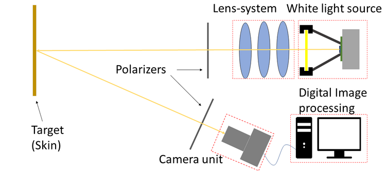
The incidence of skin cancer has constantly increased over the last years. The deadliest form of skin cancer is malignant melanoma, for which in case of early diagnosis survival rates are high. To distinguish melanoma from benign nevi certain optical criteria such as the ABCD rule or 7-Point-Checklist need to be evaluated by the physician. However, current state dermoscopy is time consuming, in particular when a multitude of skin lesions need to be inspected. As during inspection the transparent probe-plate of the dermoscope is pressed onto the skin, it flattens the lesion, thus, changing the topology. Hence, image reproducibility is difficult, in particular, for compound nevi with an out-of-plane structure. Also, the lesions at strongly curved parts of the body such as the foot are difficult to image using the standard contact techniques. In addition, for sensitive nevi and also for the diagnosis of common inflammatory skin diseases such as psoriasis, atopic eczema or Lichen ruber planus, such a contact type imaging procedure may even be painful. Therefore, within this project a versatile non-contact dermoscopic system is being developed. To obtain information from various skin depths, optical polarizers are integrated into the system. Also, a novel remote phosphor white light source with tunable color temperature was realized to additionally implement algorithms for blood- and melanin-contrast enhancement. The system also provides the possibility to inspect greater skin areas due to a large field of view. This is in particular advantageous for examination of skin diseases like psoriasis and Lichen ruber planus which usually affect larger areas of the skin. In addition, such a non-contact dermoscope can more easily be integrated into an automated medical device than its contact-type counterpart and is more suited for telemedicine applications.
In future, our device is intended to enable a fast and reliable diagnosis of malignant melanoma and inflammatory skin diseases by automated detection of suspicious skin lesions in recorded dermatoscopic images. Ultimately, such a system will significantly increase diagnostic accuracy and drastically reduce the number of lesions the physician needs to examine.
See also the Tailored Light Homepage.
