

[Text en: Raman]
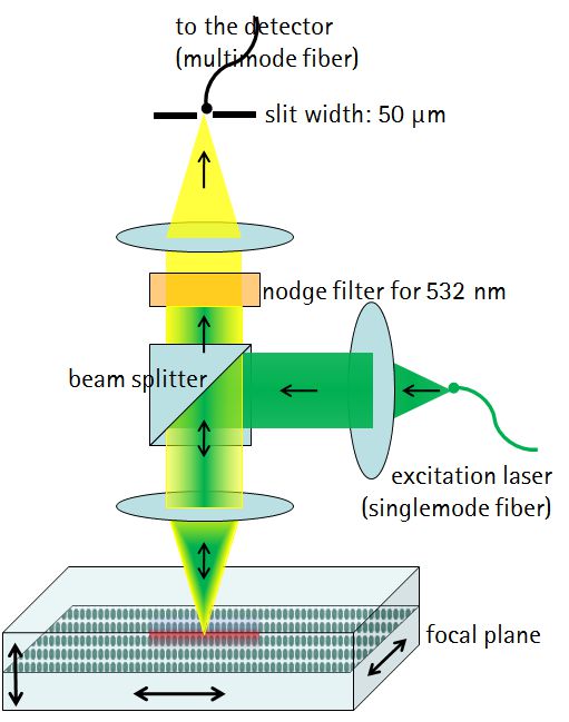
Confocal Raman microscopy combines the properties of confocal microscopy - high, spatial resolution in 3D - with the advantages of Raman spectroscopy - precise substance identification via the vibrational modes of the involved molecules.
The HOT uses confocal Raman microscopy for example to
- non-invasively analyze living biofilms
- non-invasively identify bacteria species
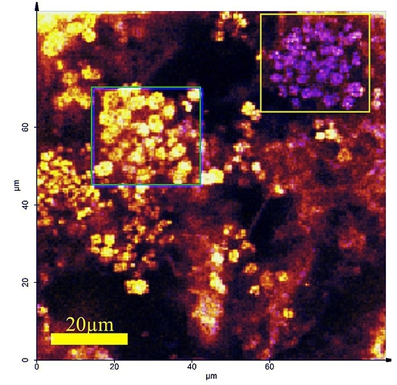
- detection of microplastics in complex environmental samples
- functional analysis of proteins, peptides, and amino acids
often using specific molecular resonances for signal enhancing or directly in the form of resonance Raman spectroscopy.
See also
- Thomas Dieing, Olaf Hollricher, Jan Toporski (Eds), Confocal Raman Microscopy, Springer 2010,
ISBN 978-3-642-12521-8, e-ISBN 978-3-642-12522-5 - Arnaud Zoubir (Edt.), Raman Imaging, Springer 2012,
ISBN 978-3-642-28251-5, e-ISBN 978-3-642-28252-2 - Pavel Matousek, Michael D. Morris (Eds.), Emerging Raman Applications and Techniques in Biomedical and Pharmaceutical Fields, Springer 2010,
ISBN 978-3-642-02648-5, e-ISBN 978-3-642-02649-2
Downloads
- Poster as PDF for Download
Confocal Resonance Raman Spectroscopy: analyzing aquaeous multispecies biofilms in vivo

